Rotator cuff tears are common. They tend to occur more often amongst people over the age of 50 than younger people in their 30s and 40s. Older people tend to get tears due to age-related changes. In fact, it’s so common almost 50% of people over 60 get them. One study showed that asymptomatic tears were twice as common as symptomatic ones! Younger and middle-aged people tend to get them due to trauma, trying to lift a very heavy object or catch something heavy. Tears can also happen from a fall onto the shoulder or arm. It can also occur from a boom-bust cycle too much too soon type of exercise such as trying a new sport and overtraining without sufficient strength and conditioning to support the shoulder. In older people, it tends to happen because of age-related changes from decreased blood supply to the tendons connecting muscles to bones.
Do I have a rotator cuff tear or a sore shoulder?
There are many conditions that can give you pain in the shoulder. Pain can be referred from the neck as well as other organs like the gall bladder, lungs, and even your heart. It’s important to know that your shoulder pain is not a symptom of any other condition. A good Physio (like all of us at Vitality Physio) will ask all the relevant questions to keep you safe.
Some of the most noteworthy features of rotator cuff tears are:
The affected shoulder hurts when lying on it while resting.
Pain when lifting or lowering the arm during simple activities such as reaching for a shelf.
A feeling that the shoulder is weak. It’s particularly noticeable when lifting your arm to shoulder level or when rotating your shoulder.
Does the rotator cuff always need surgery if it is torn?
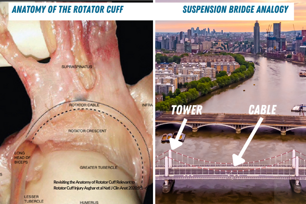
Let’s take a closer look at the anatomy:
The rotator cuff tendons are special. They are all connected via the rotator cable. Therefore, they all work together to make the rotator cuff a continuous structure, working harmoniously together. The cable functions as a stress shield. It acts in the same way that a suspension bridge transmits loads across to its supporting towers.
Here’s the good news- this means that even when you have a large tear of a rotator cuff tendon, you can sometimes neither have pain nor a loss of function!
However, tears that affect the function of the rotator cable are likely to need surgery. Therefore, the location, more than the size of the tear is more relevant in deciding whether surgery is needed.
How can Physiotherapy help?
It is important to address all factors that may have contributed to tendon injury or tear when treating rotator cuff injuries when surgery is not needed.
With physiotherapy, you will improve neck, scapular, and thoracic spine function. All of the body parts are integral to good shoulder function. If your neck is stiff or weak, your shoulder joint can often have to work harder. By targeting these supporting areas, you will improve the way your body is set up before you move your shoulder. This means that you give your shoulder the best chance to move both easily and powerfully.
As movement experts, Physios are great at distinguishing whether your joints have the stability to move efficiently too. So, we can help you get your shoulder working to its best potential.
We can help you find interim solutions to find more comfortable positions to sleep, and ways to modify your technique for activities, while you are recovering or getting stronger.
Now, generic exercises – off the internet will help to a certain extent; however, they cannot be replaced by robust, goal-orientated, and bespoke training plans. They are often not targetted to your individual needs, and it’s difficult to guess without an expert to guide you how often, how many repetitions, and how heavy to load your muscles.
To make improvements rehab should always be:
Specific- meaning is it for endurance, strength, balance, flexibility, or a combination of these?
Measurable- it needs to be scored.
Prescribed- so that you know the:
Intensity: How many repetitions?
Sets: How many times should you repeat a particular number of repetitions of a given exercise?
Dose: How many sessions per week?
By working with a well-planned specific rehab program- you’ll make the best gains and use your time efficiently too!
The shoulder is a ball and socket joint. Its structure and form mean that it allows for many different movements, making it the most mobile joint in the body! The socket is pretty shallow but this allows us not only to complete day-to-day activities like washing our hair but also allows us to participate in overhead sports like tennis and cricket. Here at Vitality Physio, shoulder injuries are one of the most common injuries that we see. But first, some anatomy:
Shoulder anatomy
Your shoulder is made up of three bones: your upper arm bone (humerus), your shoulder blade (scapula), and your collarbone (clavicle). The shoulder joint is actually where the ball of the humerus meets the rounded shallow socket of the scapula, called the glenoid. The muscles of the rotator cuff keep the large head of the humerus, positioned and centered in the glenoid. Doing so, helps to keep the most mobile joint in the body moving optimally!
So, what are the common causes of Shoulder Pain?
The shoulder is a complex joint, but fear not, with the right approach rehab needn’t be overwhelming! Overusing or underusing muscles and tendons can cause pain. Too much too soon, boom and bust are often the biggest culprits! Irritation to the bursae (the fluid-filled sac under bones or tendons) is very common and can be painful, even disturbing your sleep at night. Age-related injuries such as frozen shoulder or rotator cuff tears can cause pain too. They can rob you of movement and limit your function! Sometimes impact injuries and dislocations cause immense pain and damage to ligaments. Occasionally, shoulder pain may be referred from a nearby structure like an irritated nerve in the neck.
Every body is different and therefore every injury can present and have an impact on life in many different ways. A tendon injury for example may prevent someone from participating in sport, whereas a frozen shoulder can prevent someone from getting dressed in the morning. No matter the cause of injury, it is important to establish the cause. It helps prevent further injury, and will limit further longer-term issues too. Why leave it to chance?
What should I do if I have shoulder pain?
Distinguishing what is causing the pain, will impact your rehab. Sometimes, shoulder pain can be a result of an underlying illness such as diabetes or cardiac issues. Your Physio will flag the symptoms associated with causes other than the structures of the shoulder, and refer you to the right medical professional as needed.
If you have sudden crushing pain in your shoulder, call 999 straight away! If the pain runs from your chest to the left jaw, arm, or neck, or occurs with shortness of breath, dizziness, or sweating.
Do I need a scan of my shoulder? Why?
A thorough examination of the shoulder, alongside a detailed history of the injury, will often provide the most information required for our Physios to make a diagnosis. MRI scans are not always needed. In fact, a study in 2014 showed that MRI scans showed changes and pathology in both symptomatic and asymptomatic shoulders. It also demonstrated that symptoms may not match the MRI findings. The notable financial burden of ordering MRI scans and the pertinency of the findings are therefore sometimes questionable. This does not mean that you will never need a scan, however, this should be done to either confirm a diagnosis, rule out dangerous pathology, or if you have failed conservative therapies for a reasonable amount of time.
What can you expect from Physiotherapy?
Your Physio will offer you a tailor-made plan, based on the problems you have and the goals you’d like to achieve. Maybe you’d like to play tennis again, go bouldering or even the simple everyday activities such as getting your arm into a coat or reaching a high shelf. We are all different, so why should your treatment be exactly the same? The primary principles of our problem-solving treatments however, will all resonate with any shoulder condition that you may have. They are the mainstays of quality Physiotherapy, including education about injury, modified training, adequate strength work, restoring function, and returning safely to sport.
It’s really important to load and train the shoulder, even when pain and dysfunction are resolved. This should include a combination of strength and mobility work. Train the shoulder muscles from the famous rotator cuff, deltoids, and traps as well as the scapular and spinal muscles.
What can you do now?
If you are experiencing shoulder pain, come and see us, we will help you understand what exactly is going on. Don’t let the pain hinder you any longer. Let us help you with a tailored rehab program specific to your injury and goal.
Struggling with back pain in pregnancy? Need to know what exercises to do?
Having a baby can be joyful and daunting at the same time.
The thrills and spills of every trimester bring new challenges and new opportunities to experience the delight of motherhood.
But with all of the joys that come with pregnancy, there can also be a range of pregnancy-related physical changes and symptoms that can be tricky to overcome.
One of the most common complaints is back pain in pregnancy.
This article gives you 5 pilates-based exercises to help you ease your discomfort and feel more mobile so you can get on with your day with less pain.
How pilates exercises help for back pain in pregnancy
These low-impact exercises (no bounding or jumping) offer significant benefits, including enhancing your breathing, and helping you build and maintain strength throughout your pregnancy.
This can help prepare your body for carrying your baby and support your recovery after birth, regardless of whether you have a vaginal delivery or caesarean section.
More upsides to Pilates are that you can do it with or without equipment and it’s very versatile. So you can adapt it to your needs and abilities as you progress through your recovery.
At our clinics, we offer clinical pilates sessions tailored to your condition using props when necessary, such at the Pilates ring and Swiss ball.
Although we may use props to enhance the impact of the exercises in clinic, we also show you how to do your exercises at home without any equipment. So you can practice them as regularly as you like.
How to do these pregnancy exercises safely when you have back pain
In the video below you’ll see the top 5 exercises we recommend for pregnant patients experiencing back pain.
Before you do the exercises, it’s important to keep these precautions in mind so you don’t worsen your pain or cause further injury:
Ensure you have enough floorspace and room around you for performing the exercises.
Listen to your body; don’t push through pain. If something doesn’t feel right, take a break, or skip the exercise.
Try not to look at the screen while you’re doing the exercises. It can affect your balance and may hurt your neck. Instead, watch the demonstration then pause it. Do the exercise, then go back and continue to the next one.
Tips for getting these pregnancy back pain exercises right (and why they work)
Exercise 1: Lateral breathing
Lateral breathing will:
Enhance your joint position sense (proprioception)
Increase your lung capacity (how much air your lungs can hold)
Lengthen your spine
Condition your deep abdominal muscles (core)
Exercise 2: Thoracic rotation
Thoracic rotation is key to reducing lower back pain because it helps stabilise and strengthen your lower back muscles, which take on more and more load as your baby grows.
The trick is keeping your waist and hips facing forward, while your ribs and shoulders rotate around your central axis.
Exercise 3: Quadruped hip hinge
A crucial skill to master to prevent lower back pain and control it when it flares, is being able to move your hips without moving your pelvis.
In pregnancy, your pelvic ligaments can become more mobile as a result of hormonal and weight distribution changes. This can cause your pelvis to lose some of its support functions and resulting in lower back pain.
This exercise helps add extra support in your pelvis while moving your hips.
Exercise 4: Quadruped lateral rotation
Similarly to exercise 2, here we focus on opening the chest, whilst keeping the lower back stable.
Notice the difference between stabilising the pelvis here and during exercise 2: Which one did you find easier?
Exercise 5: Semi supine pelvic rotation
This is the most challenging exercise (that’s why it comes last).
As you shift your weight from the left to the right side of your pelvis, focus on maintaining a neutral curve in your back.
Imagine your tailbone always being the heaviest point, with a sense of lightness in the lower back.
Keeping the two sides of your pelvis aligned (without dropping one half during the rotation) helps to strengthen the pelvic ligaments and balances out the pregnancy-related changes mentioned above.
Conclusion
These exercises will help you enjoy your pregnancy more by reducing your back pain. They work by improving your posture and keeping your pelvic floor and deep abdominal muscles strong and functional well.
Things to remember:
Make sure to practice these exercises regularly so your muscles remain balanced throughout your pregnancy.
Exercises should feel comfortable and no movement should be too difficult – always modify where necessary!
If you still experience pain or the exercises aren’t getting easier after a few weeks, you may need a hands-on physiotherapy assessment and individual treatment to get to the root of your problem and heal it.
If you’re experiencing back pain after pregnancy, our Mummy MOT sessions can help you identify the causes.
Need some help getting started or progressing with these exercises?
Prefer to see a specialist physiotherapist in person to assess and treat your pain properly?
Book an appointment with us today and we’ll help you get back on your feet as soon as possible and feeling like yourself again in no time.
In our recent blog, we talked about back pain during pregnancy. Let’s take a look at how a specialist Pilates programme can support the health and good functioning of the pelvic floor in women and men, thus mitigating the risk of developing back pain and reducing it when it occurs.
What is Pilates?
Pilates is a system of training for the body and mind, based on principles created by Joseph Pilates.
By fostering a deep connection between body and mind this incredibly graceful exercise, creates a unique awareness and focus. Pilates practice leads to an intense and deeply satisfying workout, which leaves you feeling stronger, more flexible and at ease in your body.
During a Pilates session at Vitality, you use a range of small props (such as bands, balls, and weights) and equipment, including the Reformer.
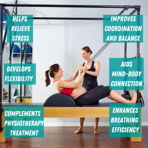
Pilates is the optimal complement to other physical rehabilitation and training programmes, such as physiotherapy, because:
It helps re-integrate the injured joint with the rest of the body.
It restores and re-trains optimal movement patterns, to prevent the recurrence of issues.
It addresses the postural factors that contribute to injury and pain.
The powerful changes that Pilates can bring
How does Pilates help pelvic floor issues?
Pilates exercises can mitigate and improve the following conditions caused by pelvic floor issues:
Back pain.
Urinary incontinence.
Anal and vaginal air.
Recovery from vaginal birth and episiotomies.
Prolapse.
Pelvic floor muscle disorder and pain.
A Pilates-based pelvic floor reconditioning programme focuses on:
Training the deep core muscles of the inner unit (transversus abdominis, multifidi, diaphragm). This means conditioning these muscles and learning how to coordinate them.
Developing efficient breathing mechanics. Optimal intra-abdominal pressure is needed to ensure the correct functioning of the pelvic floor muscles.
What does this look like in practice?
During a Pilates session at Vitality, our specialist will take you through a series of exercises to condition:
The abdominal muscles (lower abdominal fibers and transversus abdominis). For instance, you will perform exercises in a supine position where the spine is neutral and the hips move, e.g. lifting your legs up.
The back extensors. For instance, exercises in a prone or standing position where the spine is extended or stabilised against gravity, e.g. moving your limbs when in four-point kneeling.
The diaphragm. For instance, breathing exercises to practice coordinating the different muscles in the inner unit, and to practice moving in sync with different breathing patterns, e.g. the classic Pilates exercise ‘Hundred’.
How do I know if I need this?
If you are experiencing lower back pain or any other of the symptoms listed above, book your assessment with us. Our experts will determine the root cause of your issue and develop a personalised programme tailored to your needs and goals.
The third trimester promises that the fulfillment of the joys of pregnancy is imminent. Your little bundle of love is almost ready to arrive on dry land to say “hello world”. Now, the second trimester brought with it the fun days of nesting, energy, glowing skin (for some) a nice, neat bump to be proud of. Your forte was planning and organising, everything was peachy. However, with less than three months to go something seems to be going to pot. Yes, I have been there too! As a mom of two, I can totally relate to the tricky third trimester where back pain is prevalent amongst many pregnant women (every second one of us).
Back pain can be experienced in all stages of pregnancy. Often regarded as a self-limiting problem, many women are left in the dark to scrounge around the internet to find some solutions. It DOES NOT have to be this way. But first, let’s clear up a few important facts about back pain in pregnancy:
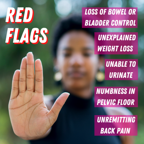
Not ALL back pain in pregnancy is self-limiting. You should see a doctor immediately if the following occurs:
You’re unable to pass urine
You have a complete loss of bowel or bladder control
Unremitting (day and night) severe back pain
Numbness in the groin, genitals, and bottom
Sudden unexplained weight loss
The above is not that common (about 2% of pregnancies). However, NEVER ignore them! They could be as a result of a concerning condition called Cauda Equina syndrome. Your spinal cord may be compressed which may result in permanent nerve damage. You should seek a surgical opinion in A&E as a matter of priority.
So, what causes back pain in pregnancy?
Back pain is complex and multidimensional, yes even in pregnancy. Our beliefs, lifestyle, habits, diet, health, sleeping, resting, exercise and emotions can influence it.
The obvious scapegoat is the cocktail of hormones to many an expectant mom in pain. I felt so guilty, when I was sick, in pain, or could not sleep at night because I was meant to be endlessly grateful for the hormones giving my growing baby all the best to expect in Chez Utero.
What do hormones do for us?
If our brain was the mothership for all human conscious and unconscious processing, our endocrine system would certainly be the first commander for all missions.The entire development of the baby from when they are simply two cells with ½ the DNA each on a mission to unite, divide, thrive, and conquer is controlled by hormones. In the early stages,
FSH (follicle stimulating hormone) is responsible for the growth of eggs in the ovaries.
hCG (Human chorionic gonadotrophin) is a hormone produced by the cells that surround the early embryo and can be detected in the urine after as little as 7-9 days after fertilisation. Over the counter pregnancy tests reveal the presence of this hormone and hence whether you are pregnant.Those cells that produce hCG go on to form the placenta.
Oestrogen
Not only is a female’s sexual development and menstrual cycle, but this hormone is also the wonder hormone that protects bones and joints. Oestrogen not only helps the uterus grow but maintains uterine lining too, but acts as an important, regulator for other hormones. Oestrogen also helps the development of baby’s organs. This powerful hormone also causes pregnant women to have a stuffy nose and causes hyperpigmentation patches on the skin.
Progesterone
Progesterone helps to regulate the menstrual cycle, and in pregnancy, helps thicken the lining of the uterus. The lining is like a soft cushion ready for a fertilised egg.The ovaries usually produce progesterone but when pregnancy occurs, higher levels are produced by the placenta.Progesterone together with, relaxin are on a common mission to prepare the mom’s anatomy for later pregnancy and eventually labour. They help soften ligaments and cartilage, and make a mom’s joints more pliable and mobile to allow for the growth of your little one.
Relaxin
I remember when I first read about this hormone as a 4th year Physiotherapy student, I was convinced it was an oversimplified and convenient name for a hormone, (akin to renaming the uterus- the womb). But no, it is indeed the scientific name and relaxin meets the full expectation of what it sounds like.Over each trimester it “relaxes” or softens your ligaments and joints in preparation for labour. However, because the hormone is released into your circulatory system, it does not only affect the pelvis but the entire body, making joints more mobile. It can sometimes cause you to feel unbalanced too. In the third trimester, relaxin also softens widens, and lengthens the cervix.
Oxytocin
Dubbed as the love hormone, oxytocin has been found to be present in higher volumes in people in the early stages of romantic attachment. The warm fuzzy feelings like trust, security, and contentment are linked to oxytocin too. In pregnancy, it is mostly known for stimulating labour contractions. Once your little baby has been born, it helps mobilise milk into the breasts and also aids with decreasing the size of the uterus.
Watch this handy video about what hormones do in pregnancy:
Biomechanics
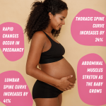
The incredible multiplication of cells is complex, beautiful, genetically predetermined. This astounding process mind-blowing to say the least is so rapid, with profound changes in ligament laxity and an enlargening uterus. Your centre of gravity moves forward, a lot! So, the lumbar spine and sacroiliac joint have to work harder to withstand these changes and increase in load. Let’s put this in context. According to Yousuf et al. 2011, the angle of the thoracic spine curvature increased by 12% between 12 and 22 weeks and increased by a total of 24% between 12 and 32 weeks. They also found that the lumbar spine curvature increased by 18% between 12 and 22 weeks and by a total of 41% between 12 and 32 weeks. The human spine is robust and responsive to change, however, these rapid changes in the spinal curvature can sometimes cause overload, and if the supporting muscles and structures are not robust and strong enough, it will cause a failure of the tissues, and can cause a strain on discs, facet joints and the sacroiliac joint too.
Similarly, as the baby grows in size, the abdominal muscles stretch and lose their effectiveness as dynamic stabilisers of the lower back too. Sometimes the muscles can separate (diastasis recti) which can cause an inefficiency in the way your back and sacroiliac joint tolerates load and can cause pain too.
Weight gain
Have you heard the urban legends and old wife’s tales before?
“Eat what you want- you’ll lose it when you give birth”
“The baby will keep you busy and the weight will fall off”
“Keep eating, the baby gets it all”
There are others too, some so ridiculous, I cannot even bear to type them!According to a 2020 study, women with a Gestational weight gain (GWG of ≥15 kg) had a higher prevalence of persistent lower back pain.But why? The reason may not be as simple or obvious. The reality is that our spines are strong, robust, and resilient and can withstand load. We will keep reminding you of this because there are far too many worry mongers out there spreading false information. In 2014, researchers found that BMI (body mass index) did contribute to back pain but did not necessarily cause structural changes in the spine. However, increases in adipose (fat) tissue may cause metabolic changes that could be responsible for back pain. In patients with a BMI over 30 overexpressed pro-inflammatory cytokines are present. Those increased levels of cytokines are considered to be the link between obesity and inflammation.
Why should you avoid obesity in pregnancy?
Elevated levels of chronic inflammation can cause and detrimentally increase leptin in the body. Leptin is a hormone in the body, secreted by fat cells that helps us regulate our body’s energy. Too much leptin will stop your hypothalamus from knowing when it is full. (The hypothalamus is in the middle of the brain and lets us know when we are full and satisfied by food, or thirsty) The more fat cells that we have the more leptin your body will make. So, too much leptin means not knowing when you are actually full! Therefore, again we can see that maintaining a healthy BMI during pregnancy will help you know when you have eaten the correct amount of food!
Stress and anxiety
Pregnancy is a life-changing life moment and can come with a rollercoaster of emotions, highs, lows, even the expected. Birth stories, the financial considerations of preparation for your little one, changing family dynamics, the impact on relationships can all set your mind racing. Everybody responds differently to stress, some of us need it to motivate us and some of us suffer negative consequences. A recent study amongst healthcare workers showed that psychological stress increased the incidence of lower back pain. There’s plenty of evidence in good-quality research about the negative effect that stress has on back pain. Pain is complex and has many dimensions and should not be oversimplified. There are so many contributing factors! Perceptions, history, and social factors also play an important part. We call this the biopsychosocial framework. A complex model demonstrates that pain is complex and multi-factorial. I prefer calling it the patient-centered approach. What we need to understand is that pain has many influences and that a variety of psychological and social factors not only affect back pain but also can impact your life, and function.
Survival of the fittest, does stress really need us to react this way?
Stress, can raise your body’s resting cortisol, stimulate, and exhaust your fight or flight system (sympathetic nervous system) and perpetuate pain. The stress associated with having a baby and the chemicals associated with that can then make it easier for future stresses to bring on pain. It becomes a bit like a trigger such as a piece of music or a fragrance that can remind you of a specific moment in your life. Inevitably over time, this can change the way the body behaves. Eventually, this can actually cause a depletion of serotonin (happy hormones).
We live in a stressful world, so what?
Stress is a normal part of life. It is not the stress that is bad but our response to that stress. If you have a big increase in stress or something has changed in your life where it’s harder to cope with stress, then stress can increase your sensitivity and can increase your chance of having pain. Not only is stress (and stress tolerance) related to the persistence of pain, it is also related to how we recover. Prominent levels of stress can impair our recovery from physically taxing components of our lives. Sometimes a stressful or traumatic event can cause us pain. That stress and the chemicals associated with that can then make it easier for future stress to trigger pain. Has a song or smell ever brought a memory back to your mind? So too can a stressful moment trigger pain. It does not mean that you are weaker or have injured or re-injured yourself! All it means is that you have a heightened sensitivity and it’s easier for normal life stressors to trigger a pain response.
Lack of sleep
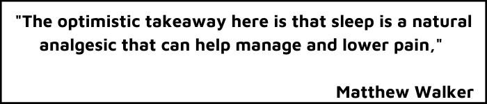
Sleep is protective. It helps us to both heal recover. A powerful desensitizer, often more potent than medicines. It builds our tolerance to all the things that can sensitize us. Lack of sleep (less than 7 hours for some) or interrupted sleep can sensitize you. So, perhaps your pain should be 1/10 but consistently missing sleep amplifies that pain to 4/10. . Lower levels of sleep have been linked with both increases in injury (e.g stress factors in athletes or the military) and increases in pain related to changes in the nervous system’s sensitivity. In his book “Why we sleep” by Matthew Walker, shares two of his sleep studies- one online and the other in a sleep lab. Walker shows how sleep deprivation and poor quality sleep affect the brain and how we perceive pain.
Does a lack of sleep make you feel grumpy?
Walker found that there was increased activity in the pain centres of the brain (the somatosensory cortex) and not to our surprise, decreased activity in the reward seeking part of the brain (nucleus accumbens) . Dopamine, the “feel-good hormone” is a neurotransmitter. It’s associated with satisfaction, pleasure, and rewards. Eating a delicious meal, winning a game or helping someone in need caused a surge of dopamine in the brain! Dopamine helps obtain goals, through the feelings of pleasure-reward and also helps us avoid unpleasant or painful stimuli. So, a lack of sleep caused decreased levels of dopamine and hence explains the misery we often feel when we lack sleep.
What is pain sensitivity and what does it do?
Another key brain region found to slow down in the sleep-deprived brain was the insula, which evaluates pain signals and prepares the body to respond. Even small changes in sleep patterns showed changes in pain sensitivity. This means that even slightly less sleep in a night showed that patients experienced higher levels of pain the next day. The self-preserving part of the brain analyses pain and picks our own body’s painkillers to kick in to help. However, a lack of sleep causes this mechanism to not work as efficiently.
It’s really important to us at Vitality Physio to debunk myths and give answers to our patients about their pain. We will empower you to understand, manage, cure, and prevent injury. Book with us for a free chat about your back pain and we can advise the best ways to help! Whichever stage of pregnancy you’re at, you can count on us to help you with your back pain.
ACL Injuries in Football: What You Need to Know — and How to Get Back on the Pitch
An ACL injury is one of the most feared diagnoses in football. The crack, the instant instability, the sinking feeling — and then the long road ahead. But here’s what we know: with the right rehab, most players return to the game they love. This article covers everything you need to understand about ACL injuries — what causes them, what recovery looks like, and what it takes to get back on the pitch safely.
What Is the ACL — and Why Does It Matter?
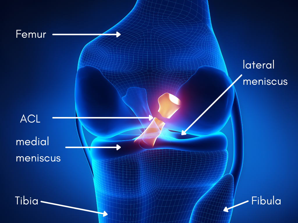
The anterior cruciate ligament (ACL) is a band of connective tissue that connects your tibia (shin bone) to your femur (thigh bone) inside the knee. It does two critical jobs.
Mechanical stability: it stops your tibia from sliding forward on your femur and controls rotational forces through the knee.
Sensory signalling: the ACL is densely packed with nerve receptors that act as a data centre for your brain, relaying real-time information about your knee’s position, tension, and movement as you run, jump, twist, and change direction.
When the ACL is damaged, you lose both. The knee feels unstable. Your brain loses the fine-grained feedback it relies on to coordinate movement. That’s why ACL rehab isn’t just about rebuilding strength — it’s about retraining the whole neuromuscular system.
How Do ACL Injuries Happen in Football?
Not how you might think. A landmark 2020 study published in the British Journal of Sports Medicine examined 134 ACL injuries in elite football players and found:
44% were non-contact injuries
44% were indirect contact injuries
Only 12% involved direct contact to the knee
That’s 88% of injuries with no direct blow to the knee. So what’s actually causing them?
The mechanics of injury
The most common triggers were:
Pressing (47%): the high-intensity movement to win the ball from an opponent
Being tackled (20%): defensive body contact while in possession
Kicking and balance (16%): maintaining stability during or after a strike
Landing from a jump (7%): particularly on one leg with rotational forces
A key finding was mechanical perturbation — when a player’s upper body is bumped or pulled while their lower body is planted on the ground, the resulting twisting force through the knee dramatically increases ACL injury risk. Pressing, tackling, and defensive actions are all high-risk moments, not because players are colliding knee-to-knee, but because of the aggressive whole-body movement interactions involved.
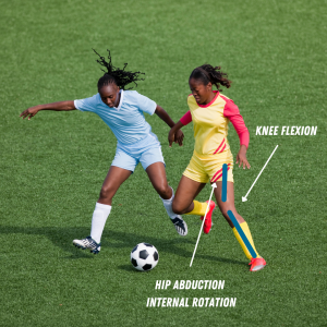
The high-risk position: dynamic knee valgus
The most common mechanism of injury is dynamic knee valgus — where the knee collapses inward as the hip rotates internally and the foot is planted. This position places enormous stress on the ACL. It’s most likely to occur during deceleration, landing, or a rapid change of direction. It can also happen subtly during pressing and tackling when the player is off-balance.
Identifying and correcting this movement pattern is a cornerstone of both ACL rehab and injury prevention.
Timing: when in the match do injuries happen?
The same 2020 BJSM study found that 25% of all ACL injuries occurred in the first 15 minutes of the match — before fatigue can be blamed. This matters because it shifts the conversation away from ‘the player was tired’ and towards readiness: how prepared is the neuromuscular system at kick-off?
Proper warm-up, cognitive readiness, and pre-match activation are not optional extras. They are injury prevention.
A Note on Female Athletes
Example of dynamic valgus load on knee.
Female footballers face a significantly higher risk of ACL injury than their male counterparts — estimates range from two to eight times higher, depending on the sport and study. Contributing factors include differences in hormonal fluctuation across the menstrual cycle, biomechanical differences in landing patterns, and neuromuscular control. At Vitality Physiotherapy, our team has specific expertise in female athlete ACL rehab and prevention — something we factor into every programme we design.
Is an ACL Tear Career-Ending?
“Be patient, don’t cut corners, and do everything the Physio tells you.” — Alan Shearer CBE
No. And there’s compelling evidence to back that up.
A widely cited study reported that 83% of athletes return to professional sport following ACL reconstruction. The key variable isn’t the injury itself — it’s the quality of the rehabilitation.
Consider Alan Shearer. On Boxing Day 1992, playing for Blackburn Rovers against Leeds United, he ruptured his ACL in the first half — and didn’t realise it. He went on to score twice in a 3-1 win. After surgery and a structured rehab programme, he returned the following season to score 31 goals in 40 games. He credits that return to one principle: do the work, follow your physio, don’t rush it.
The players who struggle to return are, more often than not, those who return too soon — before their knee is truly ready.
How Long Does ACL Rehab Take?
The standard timeframe is 9 months — but this is a minimum, not a target. Virgil van Dijk returned at 9 months after his 2020 injury. Others take 12–18 months to reach full competitive fitness. The timeline depends on:
Whether you had surgery or are managing conservatively
The extent of any associated injuries (meniscus, other ligaments)
Your pre-injury fitness level
How consistently and intelligently you complete your rehab
Your neuromuscular readiness — not just structural healing
The research is clear: returning at 9 months carries a significantly lower re-injury risk than returning at 6 months. Every week of quality rehab reduces that risk. There is no shortcut that doesn’t cost you later.
What Does ‘Ready to Return’ Actually Mean?
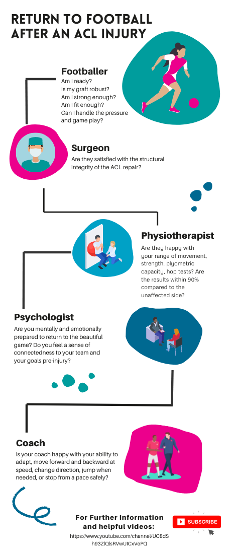
Return to football infographic by Vitality Physiotherapy
Return-to-play is not a date on a calendar. It’s a decision made by a team — athlete, physiotherapist, surgeon, coach, and sometimes a sports psychologist — using objective data. Here’s what that looks like in practice.
The athlete
Do you trust your knee? Can you commit to a tackle, a sprint, a jump — without hesitation or fear? Psychological readiness is not a nice-to-have. Fear of re-injury is one of the strongest predictors of not returning to pre-injury performance levels. Your inner voice matters as much as your single-leg hop score.
The physiotherapist
Your physio needs to see objective evidence of readiness across multiple domains:
Strength and power: can you achieve 90%+ limb symmetry on strength testing?
Hop testing battery: single leg vertical hop, forward hop, timed 6m hop, triple hop, triple crossover hop, lateral hop, medial hop, and their rotational variants. This isn’t an arbitrary list — each test assesses a different demand of football movement.
Neuromuscular control: reaction times, adaptability under fatigue, dynamic balance
Movement quality: can you decelerate, change direction, and land without compensating?
The surgeon
Structural integrity matters. Your surgeon will assess graft healing, range of movement, and whether the reconstructed ACL has the mechanical properties required for full sport. A happy surgeon means a structurally sound knee — but structural soundness alone is not clearance to play.
The coach
Pitch-based performance has to match the test results. Can you accelerate and decelerate at full speed? Change direction under pressure? Jump and land when required? React to a ball coming at you unpredictably? Return to training is the bridge between rehab and competition — it shouldn’t be skipped.
The role of cognitive training
A 2021 study found that cognitive skills, reaction time, and processing speed are significant factors in preventing ACL re-injury. This is why progressive, evidence-based ACL rehab includes reactive drills — small-sided games, decision-making under pressure, gates games, possession exercises. The nervous system needs to be retrained, not just the muscles.
Physical fitness without cognitive and neuromuscular readiness is a re-injury waiting to happen.
The Final Whistle
An ACL tear is not relegation. It is a significant injury — but it is one that, with the right support and the right approach, most players recover from fully.
The players who come back stronger are the ones who take the rehab seriously, trust the process, and resist the temptation to rush. There are no shortcuts — but there is a clear path.
At Vitality Physiotherapy, we have the clinical experience, the protocols, and the team to take you from injury to full return to football — whether you’re playing Sunday league or semi-professionally. We work across our Southwark (SE1) and Esher (KT10) clinics, and we’d love to support your recovery.
Want to talk through your injury? Book a free 15-minute call with one of our ACL specialists — from the convenience of home.
Call us: 020 7193 9928
Email: info@vitality-physio.co.uk
References
Della Villa F et al. (2020). Football ACL injuries reloaded. British Journal of Sports Medicine Blog.
Ardern CL et al. (2018). Return to sport following ACL reconstruction. British Journal of Sports Medicine.
Grooms D et al. (2021). Cognitive and neurological contributors to ACL re-injury. Journal of Orthopaedic & Sports Physical Therapy.
We've all come across a friend or famous athlete who has had their season cut short through an ill-timed knee twist and alongside the dreaded "popping" sound. When this happens, it can be daunting.
Understanding ACL injury and knowing what steps to take to recover will help you get back to the sport you love sooner.As mentioned in our ACL tear article, the ACL is one of the main ligaments controlling the stability of your knee, and injuries to the ACL occur more frequently in sports involving landing and pivoting actions like football, netball, or skiing.
Diagnosis of ACL Injuries
The Lachman Test
John Lachman (1919-2007) was an orthopaedic surgeon at Temple University in Philadelphia who invented the technique. It's considered the most reliable way to diagnose an ACL injury. It is used by Physios and Surgeons the world over.
How is the Lachman test performed?
The Lachman test is performed like this:
Lie flat on your back. Relaxed, with legs straight.
Your Physio places your knee in a slightly bent (and sometimes rotated) position.
One hand is on the back of your lower leg (just under your knee joint), and the other is on the front of your thigh.
They then pull your shin forwards firmly while stabilising your thigh.
Why do we do the Lachman test?
To measure the endpoint and joint laxity.
The endpoint is where the end of the joint movement is felt as the shin is pulled forward. If this endpoint feels further away than what's expected, then this may indicate an injured ACL.
Joint laxity is the general feel of your joint movement and the sense of how lax (or loose) your ACL feels compared to your other knee.
Grading ACL tears with the Lachman test:
Normal. There's no difference in movement or laxity between your left and right knees.
Mild (grade 1). There's slightly more (about 2-5mm) movement than in your other knee.
Moderate (grade 2). There's moderately more (about 5 to 10 mm) movement than in your other knee.
Severe (grade 3). There's much more (10 to 15 mm) movement than in your other knee.
Because we're all different and have varying baselines of "stiffness", the test should be done on your other leg too. This will give the Physio an accurate idea of what's typical for your joint.
Anatomy of the L knee showing a ruptured ACL
The Pivot Shift test
How is the pivot shift test performed?
1. Lie flat on your back. Relaxed, with legs straight.
2. The Physio places one hand on your heel and the other just below the outer side of your knee.
3. They apply pressure to the outside of your knee while internally rotating your lower leg and bending and straightening your knee.
The test is positive if your lower leg sinks backward when the knee is bent at 30-40 degrees. Sometimes a 'clunk' can be heard when this happens.
Treatment options for ACL tears
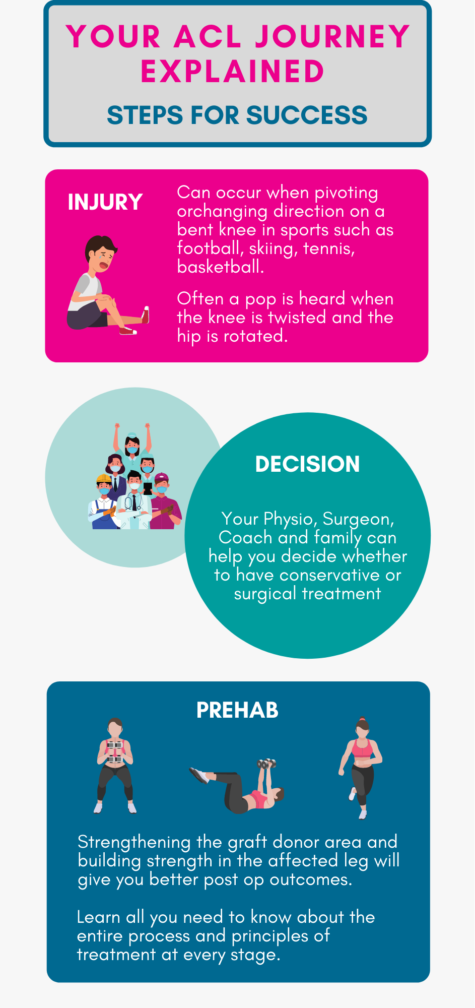
There are two treatment options for an ACL injury: Conservative or surgical management.
Your age, previous injury, and fitness and rehab goals should be considered when deciding the best route to follow.
Conservative management
Conservative or non-surgical management involves a period of Physiotherapy treatment to restore your knee function to pre-injury level.
Surgical management
Having ACL surgery usually requires a minimum of 9 months of rehab tailored towards getting you back into your sport. This may sound like a long time, but returning to sport too early can have negative implications. Mainly increasing your risk of re-injury.
ACL Surgery
What surgery is there for ACL tears, and what you can expect?
The most common type of ACL surgery is a reconstruction of a new tendon called a graft. This is typically a minimally invasive procedure done via keyhole surgery using an arthroscope (a small camera through which your surgeon passes tiny surgical instruments). First, the damaged ACL is removed, and then the graft is attached in its place. There are different types of grafts. They include:
Autografts - Formed from tissues taken from another part of your body (like tendon tissue strands from your hamstring or patella tendon)
Allograft - Formed from tissues taken from another person's body
Synthetic - Formed from an artificial source or donor.
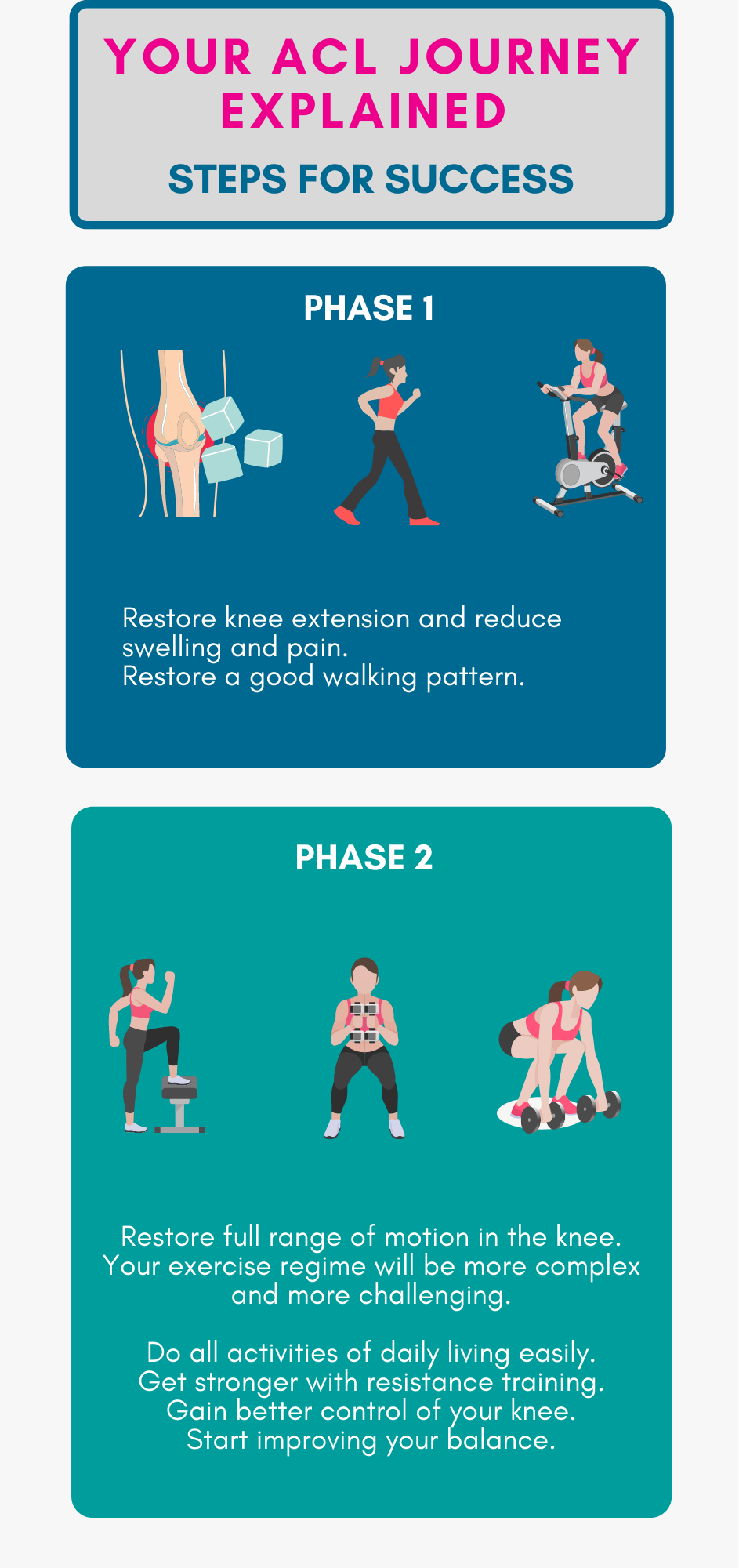
Rehab is paramount to your ACL recovery (with or without surgery) . Without rehab, ACL surgery is meaningless. All the top knee surgeons agree. Your ACL rehab determines the success of your recovery. We recommend an evidence-based programme that's divided into four progressive phases. In our experience, this process takes 9 to 18 months to complete.
Phase 1: Restore knee extension and reduce swelling
Immediately after sustaining your injury or after your surgery, the muscles around your knee may feel weak, and your joint may be swollen and painful. In addition, you may not be able to completely straighten your knee. During this stage of your recovery, the aim is to reduce swelling and pain and get your knee completely straight again. This phase typically takes 2-3 weeks.
Phase 2: Strength & Neuromuscular control
After you've passed the criteria to move onto phase 2, the fun stuff begins! You'll introduce a new set of exercises, including squats, lunges, leg presses, and more. These will continue throughout your rehab programme, becoming more complex as you advance through the various stages.
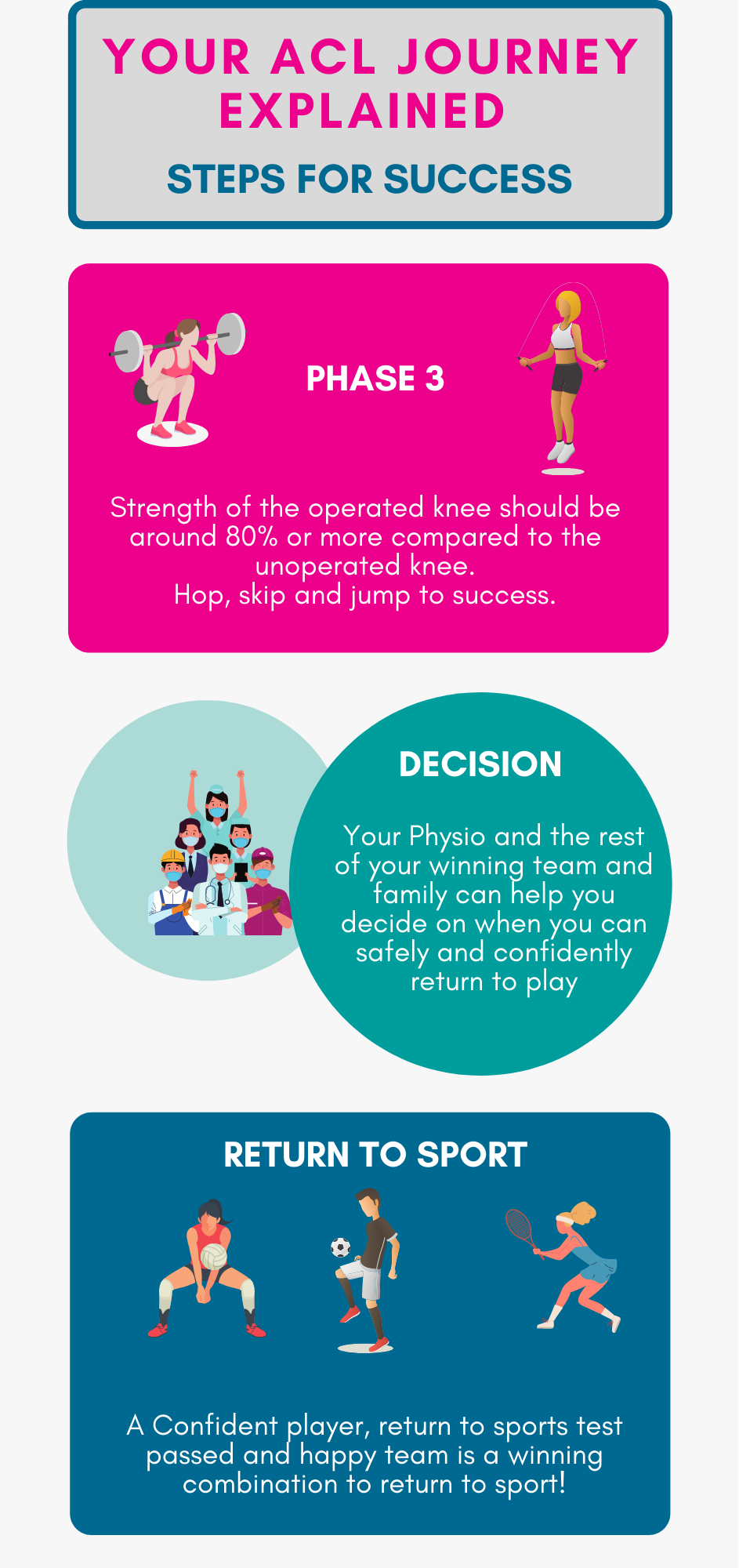
Phase 3: Plyometrics
After passing all the phase 2 criteria, you can ramp up the fun a little more and start introducing hopping, jumping, landing, and running movements. These sport-specific functional activities need to be retrained for your to safely return to your sport.
Phase 4: Return to Sport
Until this moment you have been working towards Phase 1-3. Before you get back to your sport, you and your Physiotherapist should discuss how and plan how you will do it.
General guidelines for ACL rehab
Follow each phase-specific exercise and test and only move on to the next phase once you have mastered the current one.
You need to achieve your rehab goals in the correct order to ensure you fully recover.
If you stop or skip steps in your rehab, you can worsen your current injury or increase your chances of re-injury when you eventually return to your sport.
Conclusion
Now that you know how to get a diagnosis, choose the proper treatment, and understand the rehab process better, you can move on from your ACL injury in the right direction.Yes, the rehabilitation process is long and requires dedication and patience. But, with the right team supporting you, your progress can be smoother, and it can be a whole lot more fun!
Our highly experienced and knowledgeable Physiotherapists can get you where you want to be. So, call us now and put your ACL recovery planning in our hands!
Knee pain is common. For some, the simple activities of daily living such as walking or climbing stairs can become cumbersome when your knee hurts. For others a niggle in the knee can deteriorate and adversely affect sporting performance. There are many causes of knee pain, so it is important for you to know what is causing the pain. There are however some common themes that occur with most types of knee pain.
Why does my knee hurt?
The knee is comprised of many structures including ligaments (connects bones together) tendons (attach muscles to bone) and cartilage (the connective tissue between bones). Most injuries in this area are caused by multiple problems over a long period of time- this could include muscular weakness, overuse, direct trauma or even lifestyle factors.
Most patients say that when their knee hurts, they tend to want to do less activity as more activity requires bearing extra load through the knees. Otherwise simple activities such as climbing the stairs or even going on a run can feel scary. Often, it leads to us searching for answers by having x-rays or scans. Sometimes a scan can look really scary, and sometimes we are told that we have “bone rubbing on bone” or our cartilage has “completely gone”. This can increase fear and further reduce activity and weight bearing through the knee.
However, just like we all get wrinkles or grey hair, age related chages to our knees are inevitable. Researchers now believe that inactivity is a major contributor to age-related diseases and disabilities, and that regular exercise can reduce or reverse those risks.
What should I do if I have knee pain?
Most knee pain can be treated through a graded rehab programme. Typically, this consists of a combination of strength training, education and a good understanding of how to implement your programme. It is important to have a supervised programme so that you know exactly where to start and know exactly how you are going to progress over the course of the rehab programme.
When we strengthen the muscle around our knees we increase our knee’s ability to withstand the load that we tend to put on them on a daily basis.
FUN FACT: Forces transmitted across the knee joint during normal walking range between 2 and 3 times body weight!
This makes them much less likely to experience pain in the future. Over recent times we have seen a huge body of evidence to suggest that an 8-12 week exercise programme can significantly reduce knee pain and symptom progression.
What Should I Do Now?
Seek out advice from a healthcare professional who understands knee pain and is up to date with the current research.
As mentioned before, scans are not always needed, and scans often do not correlate with the pain you are experiencing.
Begin strength training with the supervision of a healthcare professional.
Other factors to consider are sleep, diet and body weight- all of which can contribute to pain and pain sensitivity.
Here are some great simple knee exercises to try at home:
The ten most common symptoms of Long COVID were highlighted as fatigue, shortness of breath, muscle pain, cough, headache, joint pain, chest pain, altered smell, diarrhoea, and altered taste.
The two main symptom clusters for people experiencing Long COVID were:
fatigue, headache, and upper respiratory complaints.
ongoing fever and gastroenterological symptoms.
What is fatigue?
Fatigue is a feeling of constant tiredness or weakness, that does not improve with rest. Despite the fact that fatigue is sometimes referred to as tiredness, fatigue is not the same as being tired or sleepy.
A person often will feel fatigued for a number of reasons, whether it is physical, mental, or a combination of both. People of all ages will experience fatigue at some point in their lives.
In the case of viral infections such as COVID-19, fatigue is a normal part of the body’s response. After an infection is cleared, fatigue sometimes persists for some time. Fatigue can make you sleep more, feel unsteady on your feet, it can make exercise very difficult and affect your ability to concentrate and remember.
What causes post-viral or Long covid fatigue?
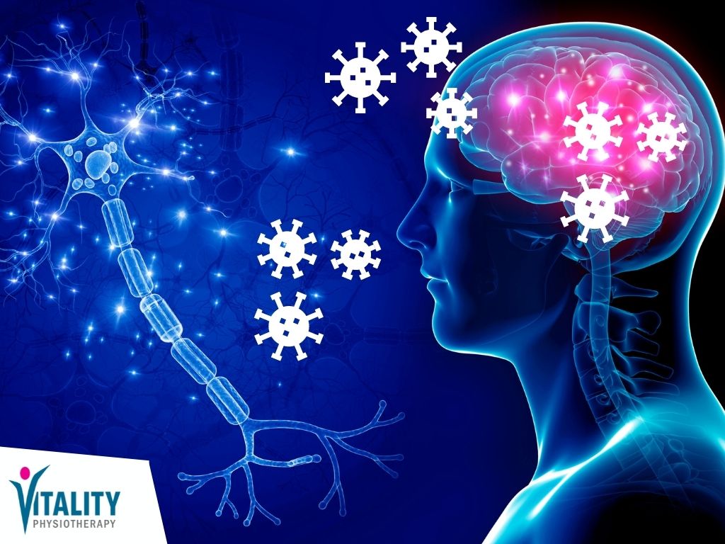
We now think that (based on post-mortem SARS research), the virus crosses the blood brain barrier.
Proposed Mechanism of how Long Covid causes fatigue by Vitality Physiotherapy
The blood-brain barrier is a specialised boundary between the blood supplying the brain and the spaces between the cells of the brain (the extracellular matrix). This incredibly specialised feature of the brain is very sensitive and selective. It allows very few substances to move from the blood into the brain, stopping toxins and harmful substances from entering the brain. It allows useful substances such as water, glucose, and oxygen to enter the brain. For more detailed information on this important mechanism, watch this:
The current idea about how the virus probably enters the brain is that when the virus enters your nose, it travels through the olfactory (nasal pathway). Then, it travels through the soft tissue in the nose via the drainage mechanism of the brain (lymphatic system). It may also possibly explain why anosmia (lack of ability to smell) is observed in patients with COVID-19.
Pro-inflammatory substances, such as cytokines (interferon gamma, and interleukin 7) start to increase in the brain area. This seems to affect the glymphatic system (macroscopic waste clearance system that use tunnels around blood vessels, formed by astroglial cells (star-shaped nerve cells), to help rid the brain of soluble proteins, waste, and metabolites from the central nervous system. The glymphatic system (largely active whilst we sleep) also helps deliver glucose, lipids (fats), proteins, growth factors, and neuromodulators to the brain. This is one of the reasons why we as humans need quality sleep because many of the toxins in our brains are eliminated when we do so.
The cytokines likely pass through the blood-brain barrier in organs such as the hypothalamus, leading in the longer term to interruption of the sleep/wake cycle, cognitive dysfunction and profound fatigue (post-exertional malaise), all characteristic of Chronic Fatigue Syndrome Myalgic Encephalomyelitis (CFS/ME).
We think that as it was the case with the SARS outbreak, some patients who contracted the COVID-19 virus may go on to develop a severe post-viral syndrome we term ‘Post COVID-19 Syndrome’ or Long COVID
What can I do to help myself?
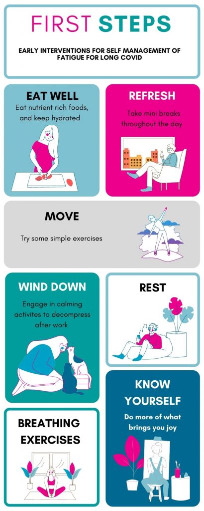
There are many interventions and self-management strategies to help with ongoing fatigue after recovering from your initial infection.
Nutrition
Eat regular healthy nutrient-rich foods including fresh vegetables, fruit, carbohydrates, and healthy fats. Vitamin D has been shown to decrease viral load and boost your immune system, so get some sun too! Remember that your body is comprised of between 50 and 65% water and it is crucial to your recovery to keep topped up.
Refresh
A short break every few hours is so beneficial to enhance your wellbeing and productivity. A mini-break does not need to be complicated. It can simply be a few minutes away from your work; this may be chatting with someone who is in the room with you or getting a drink, looking at a piece of art, listening to music, doodling, or drawing.
Sleep
Sleep is critical to your recovery because when you sleep your immune system works more efficiently. Our sleep hygiene top tips are to ensure you train your body to get to sleep every night at the same time, keep your room as dark as possible, avoid caffeine after 2 pm and stop using screens at least an hour before bed.
Move
Get active, initially, it might just be a gentle 5 min stroll or a few simple arm exercises, but you can slowly build up your activity levels. A little exercise often, allowing periods of rest in between activities is a great way to start. If you’re back at work, try standing up from your desk, if you’re office-based, every hour and march on the spot. Try going for a walk every day, starting with 5 min or so, and gradually increasing your walk time
If however, you find that simple exercises like climbing up a single flight of stairs, leaves you breathless, speak to your GP. If you feel like after exercise, you experience exceptional fatigue and malaise, speak to one of our Physios, about a more structured exercise plan, that may involve goal setting, exercise tolerance prescription, and pacing.
Wind down, allow time
The severity of the virus is not directly proportional to the level of fatigue you may be experiencing. We are all ‘wired’ differently, and our experience of illness is not the same. There is no carbon copy reference, so don’t rush with your rehab. Some patients experience levels of anxiety, fear, or even sadness. Decompress your thoughts by engaging in calming activities such as meditation, prayer, relaxing with a pet, or listening to music. Activating your creative brain too, or by being “inflow” has been shown to help many people with their levels of happiness. Drawing, writing, and other repetitive creative activities activate “flow” and produce dopamine, which is a feel-good chemical that energises and motivates you.
Breathing exercises
Many of us over breathe, taking more oxygen than we require and in turn, can experience chronic fatigue. Mouth breathing is one of the biggest culprits that cause us to over breathe. Nasal breathing, on the other hand, is a more effective, more efficient method of breathing. Its benefits are vast in number and include improved lung volumes, reduced exposure to foreign substances, improve the function of your diaphragm, improve athletic performance, sleep better, and have better-smelling breath!
Check out this video about nasal breathing by our in-house respiratory Physiotherapist, Janine Enoch:
Finally,
If you are experiencing fatigue with exercise, diminished tolerance for exercise, or simply confused about where to start, speak to one of our Physiotherapists for a free 15 min chat, which we can do over the phone or in person. Help is at hand, live well with Vitality Physiotherapy.
Without it, life cannot be sustained, it is as simple as that. Breathe in, breathe out, our simple but most important automatic reflex-controlled function. Most of the time, our brain and reflexes do all the work for us. Have you ever observed a sleeping baby, breathing gently as their chest rises and falls, effortless and without any conscious process involved?
When the demand arises, we can consciously decrease or increase our rate of breathing. Do you remember the days of “hide and seek” when the suspicious seeker was dangerously so close you’d dare not breathe, or the exhilaration associated with the rapid preparatory breathes I take before that first jump off the highboard at the local swimming pool?
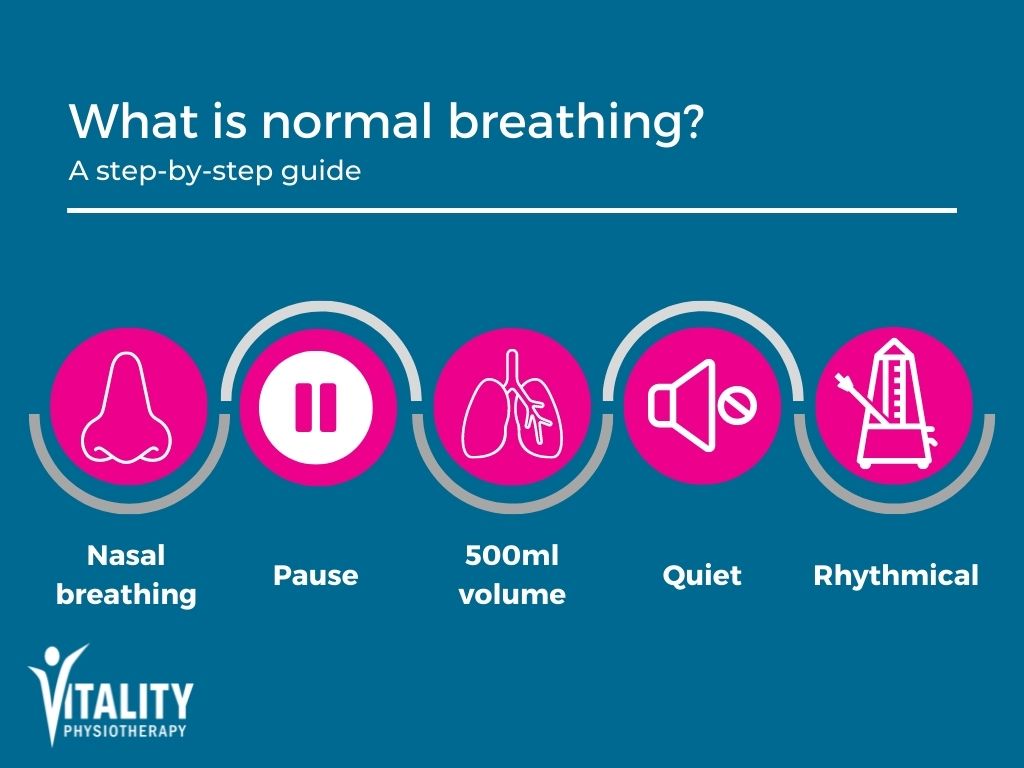
What is a normal breathing pattern?
Gentle breathing through the nose
Breathing in for 1-1.5 sec
Breathing out for 1.5-2 sec
Pause between in and out breath
Take 10-14 breaths per minute in adults
Breathe about 500ml of air per breath
Breathing is quiet
The normal breath cycle
Sometimes it goes wrong and our breathing becomes dysfunctional. What is dysfunctional breathing (DB)?
This can occur with heavy exercise, strong smells, cold weather, stress or other triggers. People would breathe rapidly through the mouth, breathe using the upper chest. The accessory neck muscles work hard and you will effectively hyperventilate.
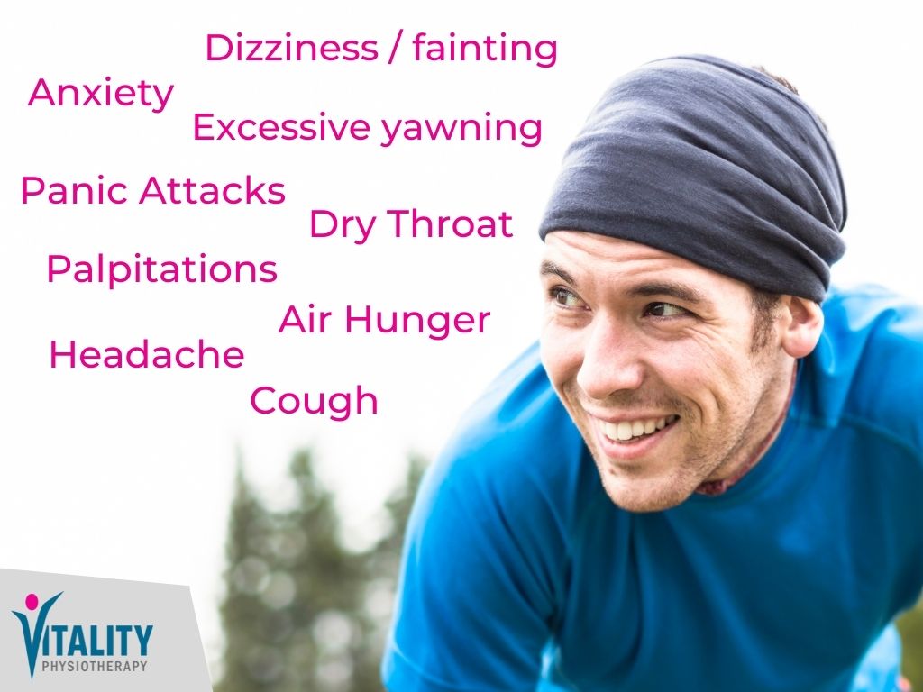
The primary symptom is often breathlessness but is usually clarified as a feeling or need for more air or “air hunger”. Additionally, DB may cause non-respiratory symptoms such as dizziness and palpitations. It has been identified across all ages. In the United Kingdom, its prevalence is approximately 9.5% among adults.
What happens when we breathe?
We breathe in air containing a mixture of oxygen (O2) and carbon dioxide (CO2) and other gases. Our bodies process and use some of the inhaled oxygen to make energy, and creates carbon dioxide as a result. When you breathe out the air contains less oxygen and more carbon dioxide. However, carbon dioxide is not just a waste product of our body’s processes, it also has a vital role in regulating the pH of the blood. During stressful situations, a higher level of CO2 in your blood, helps your body to produce adrenaline which will help you flee from danger.
How does hyperventilation affect our bodies?
Some of the most common symptoms of hyperventilation
When we hyperventiltate, and there is less CO2 and more O2 in the blood, this causes respiratory alkalosis, or decreased acidity of the blood. This causes our blood vessels to constrict and causes reduced blood supply to the brain, among other things. This causes a feeling of light-headedness and pins and needles in the fingers, muscle cramps and exhaustion.
When overbreathing becomes regular and more permanent, the brain recognizes this lower level of carbon dioxide and accepts it as being normal. Consequently, the body is constantly on alert. This cycle of hyperventilation causes a self-perpetuating cycle, that fuels back into the system making you feel more anxious, more breathless and more hungry for air.
We also alter our posture significantly when we over breathe, adopting hunched-up shoulders as a result of rapid breathing. This causes us to use our neck accessory muscles to help draw air into our lungs. Prolonged and ongoing use of these neck muscles can cause an aching neck and stiff shoulders. Typically we can overuse sternocleidomastoid and scalene muscles which are at the front of our necks to do so. Check out this helpful video to hear more about breathing pattern disorder.


 Proposed Mechanism of how Long Covid causes fatigue by Vitality Physiotherapy